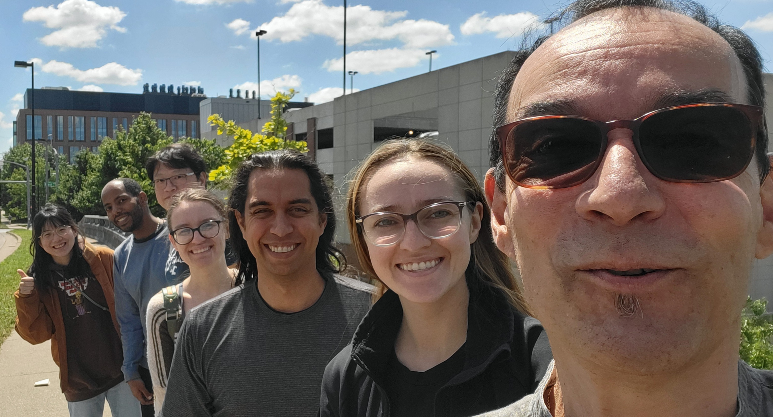
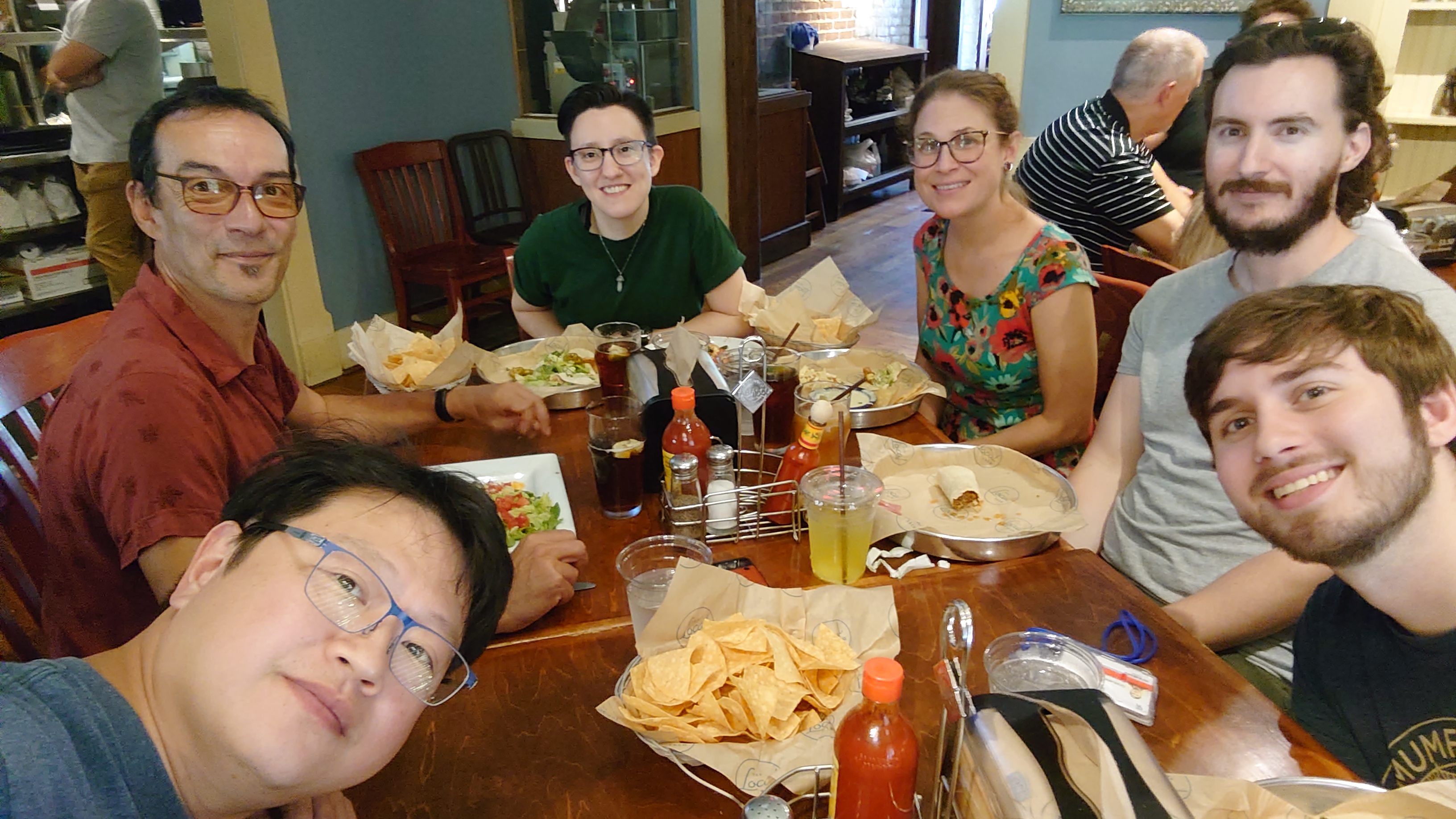
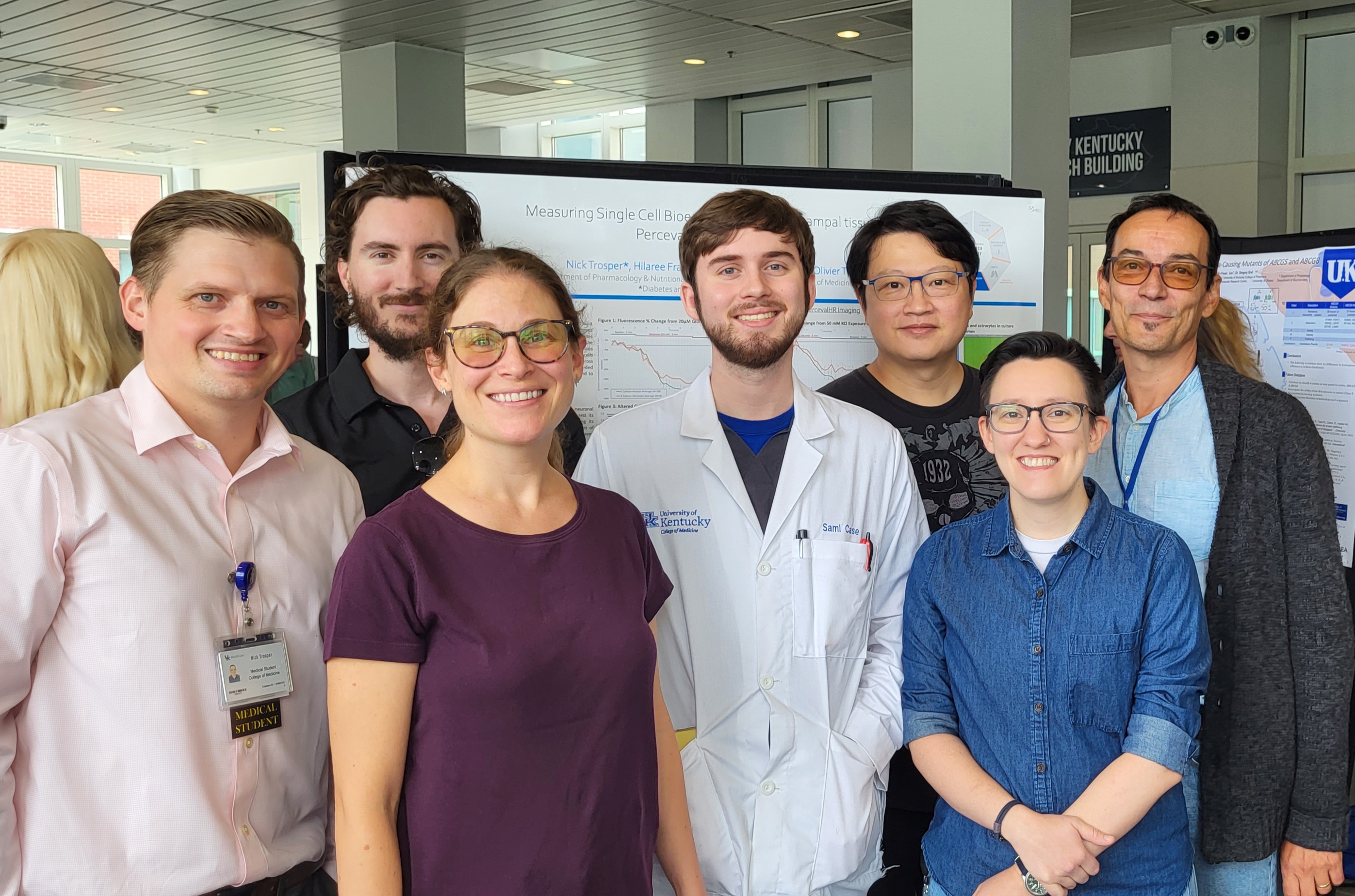
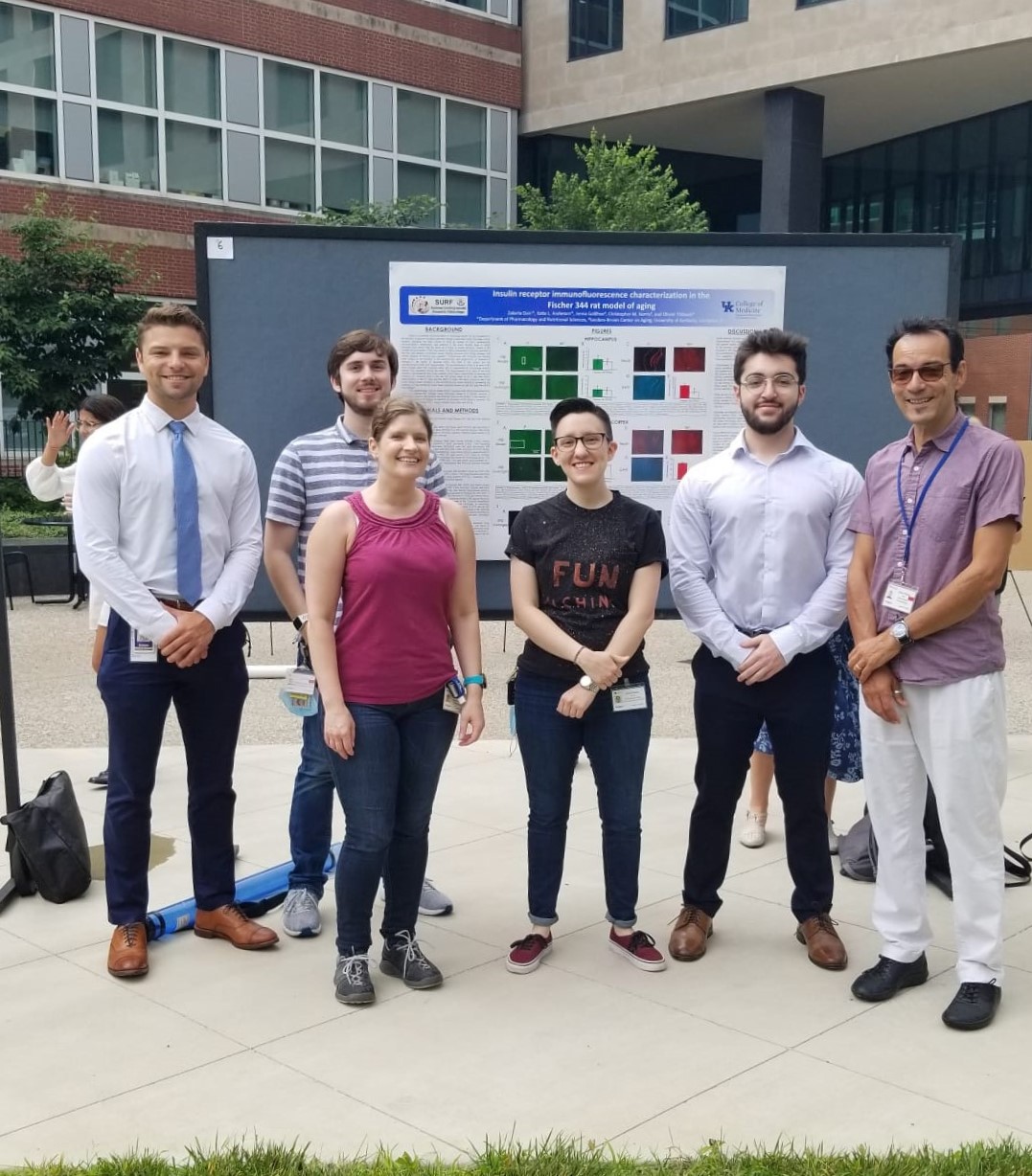
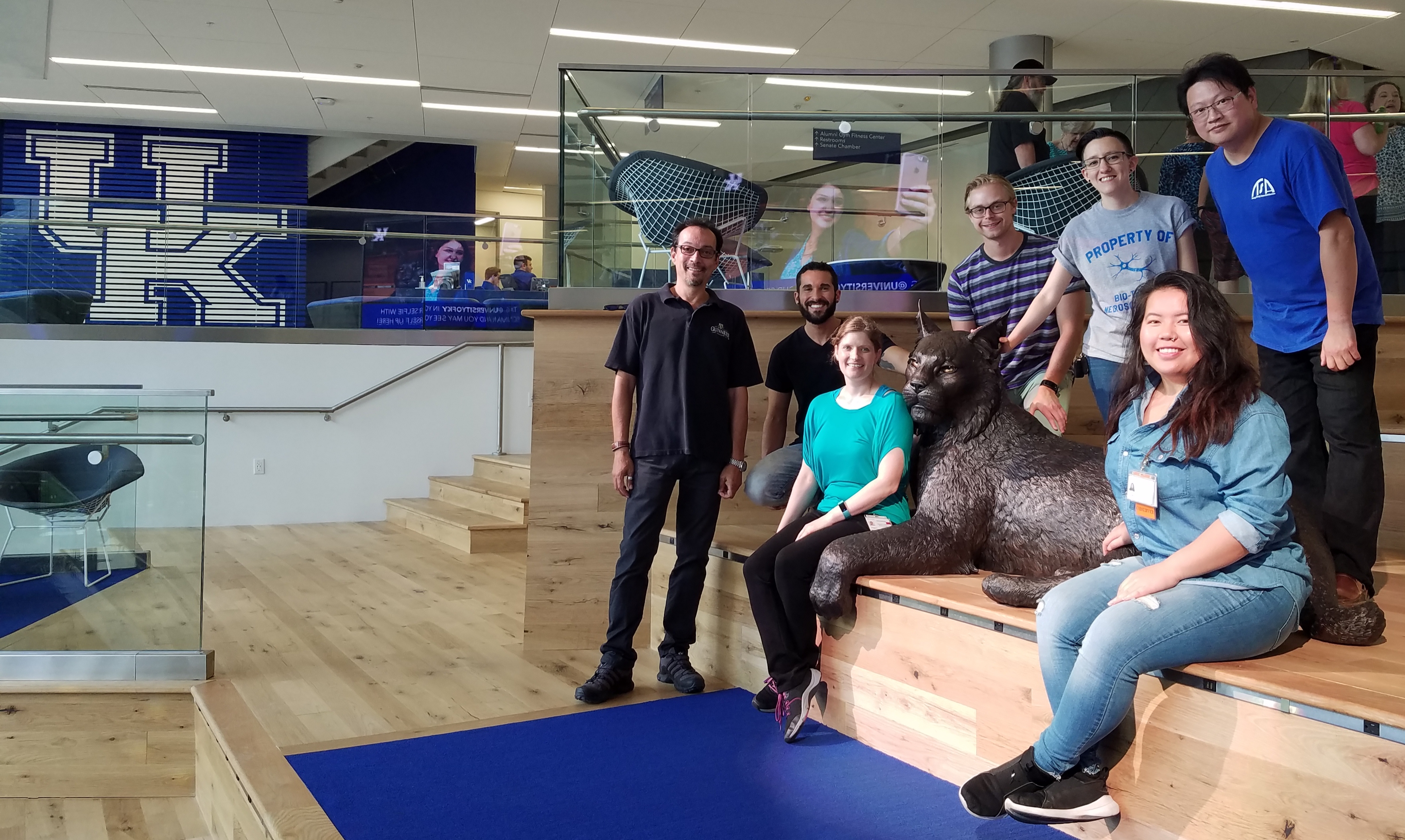
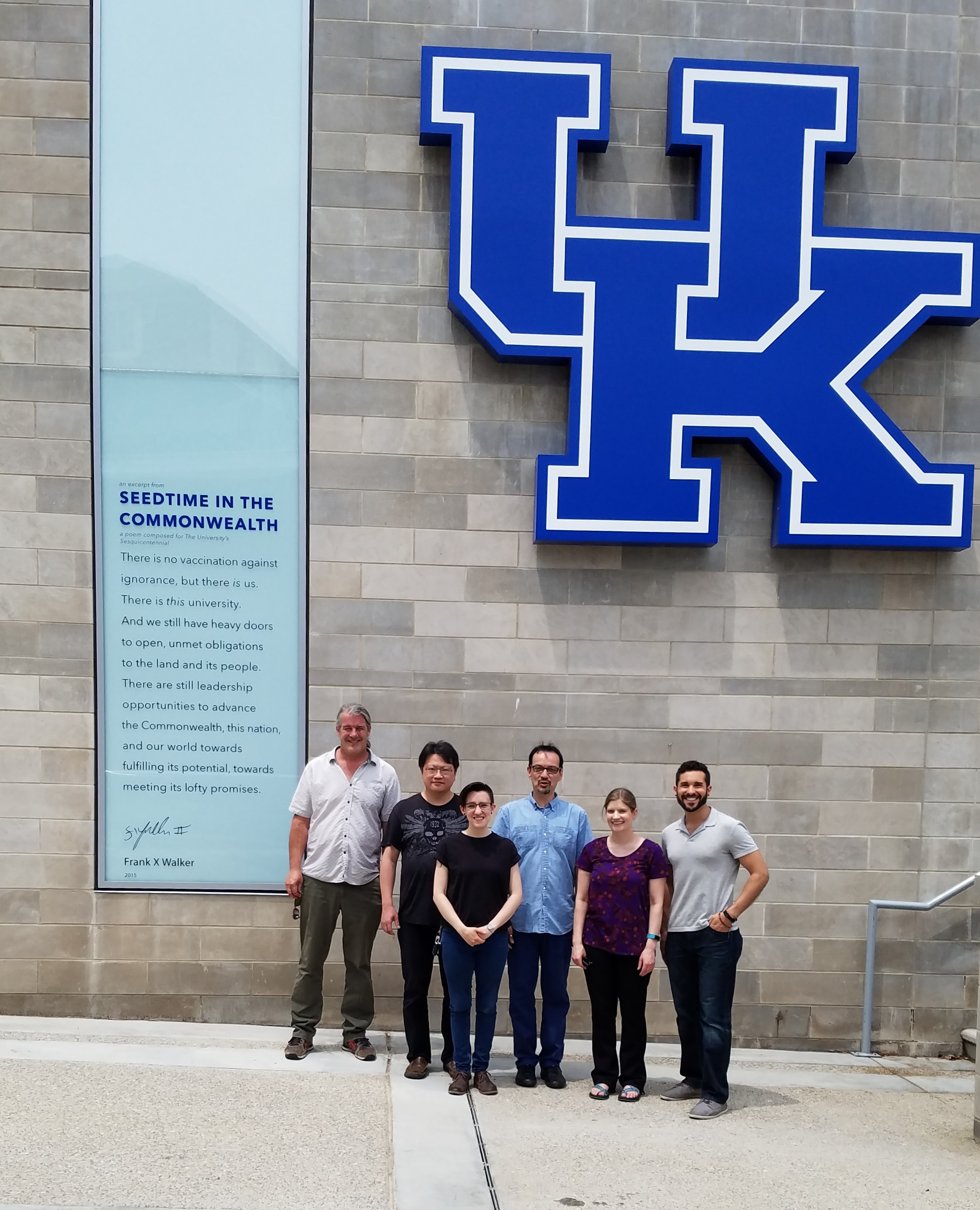
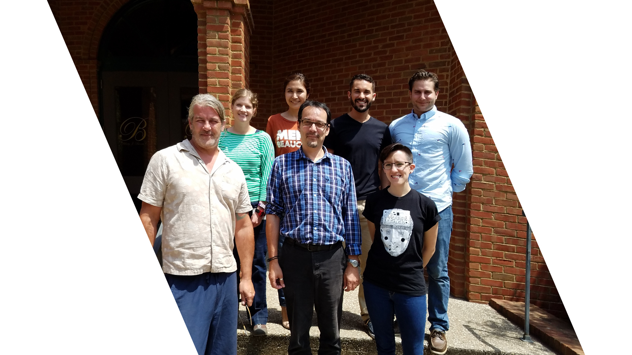
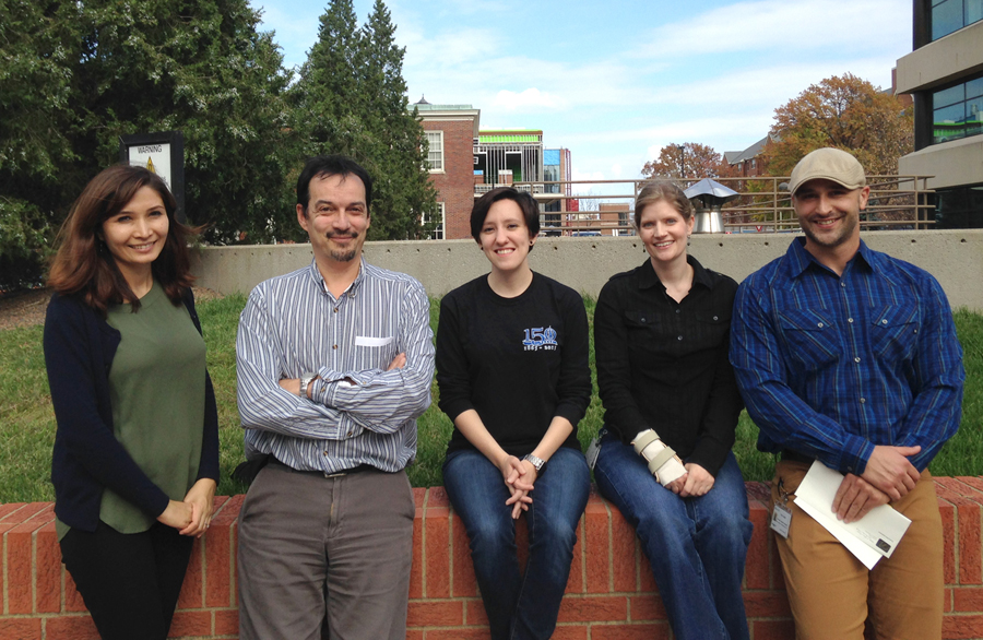
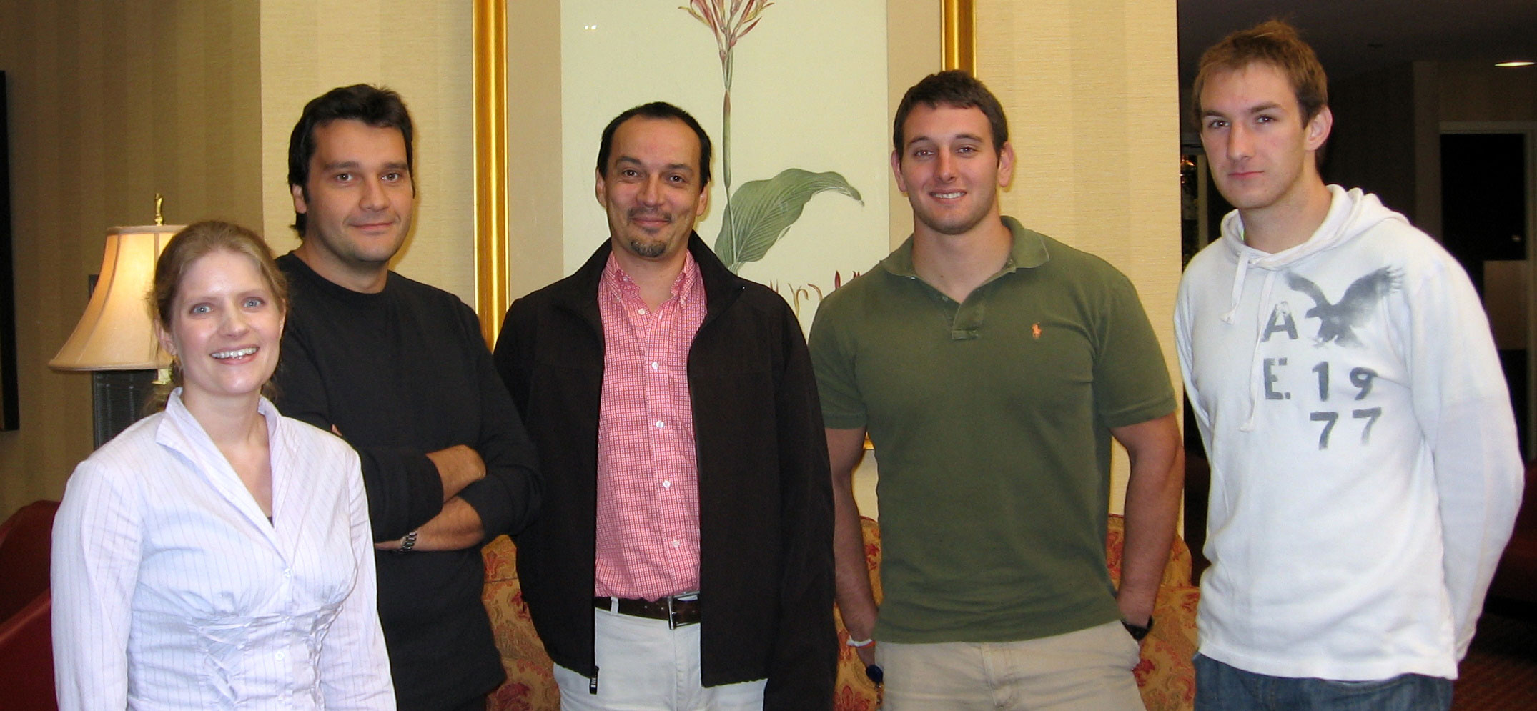
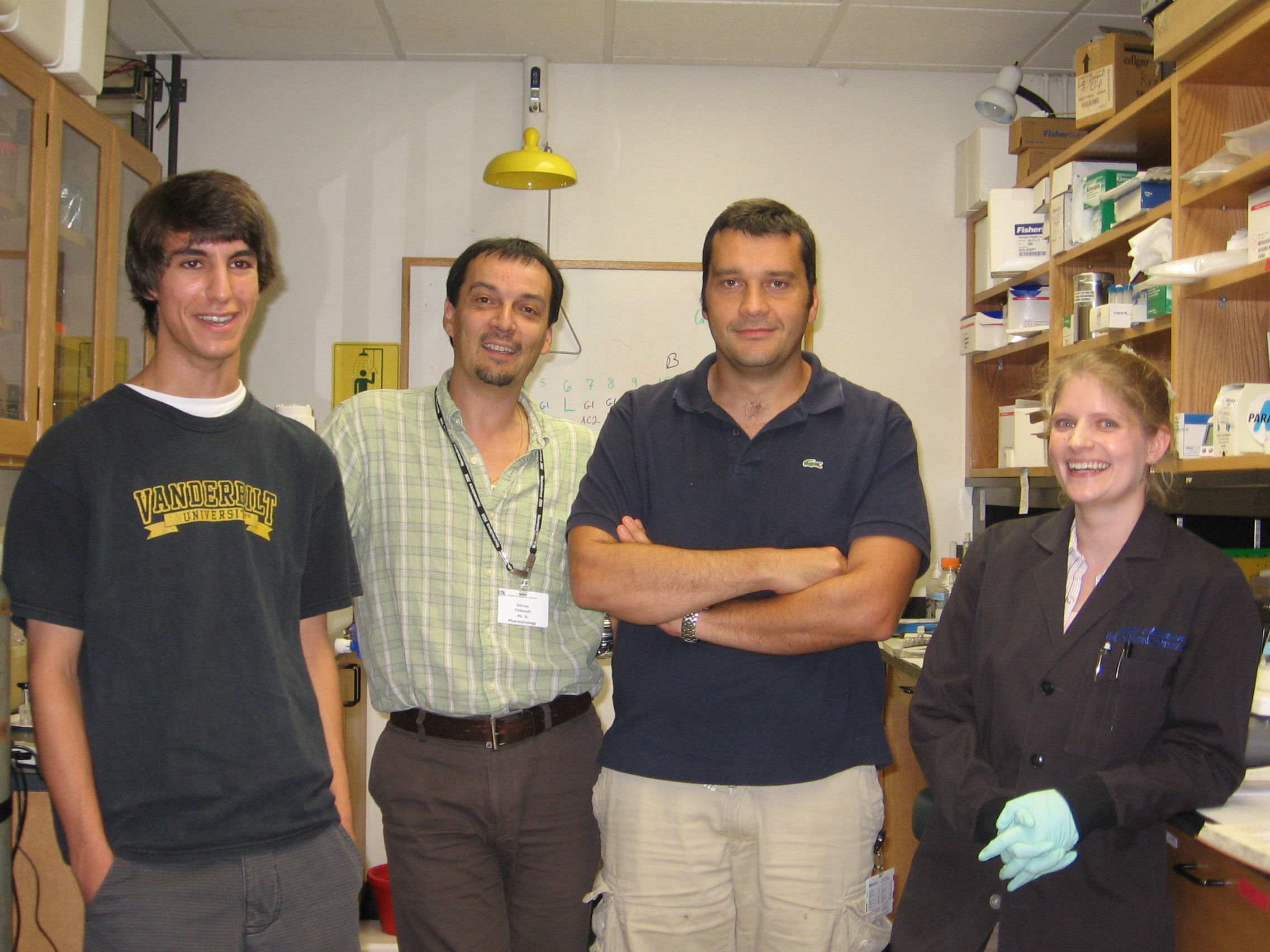
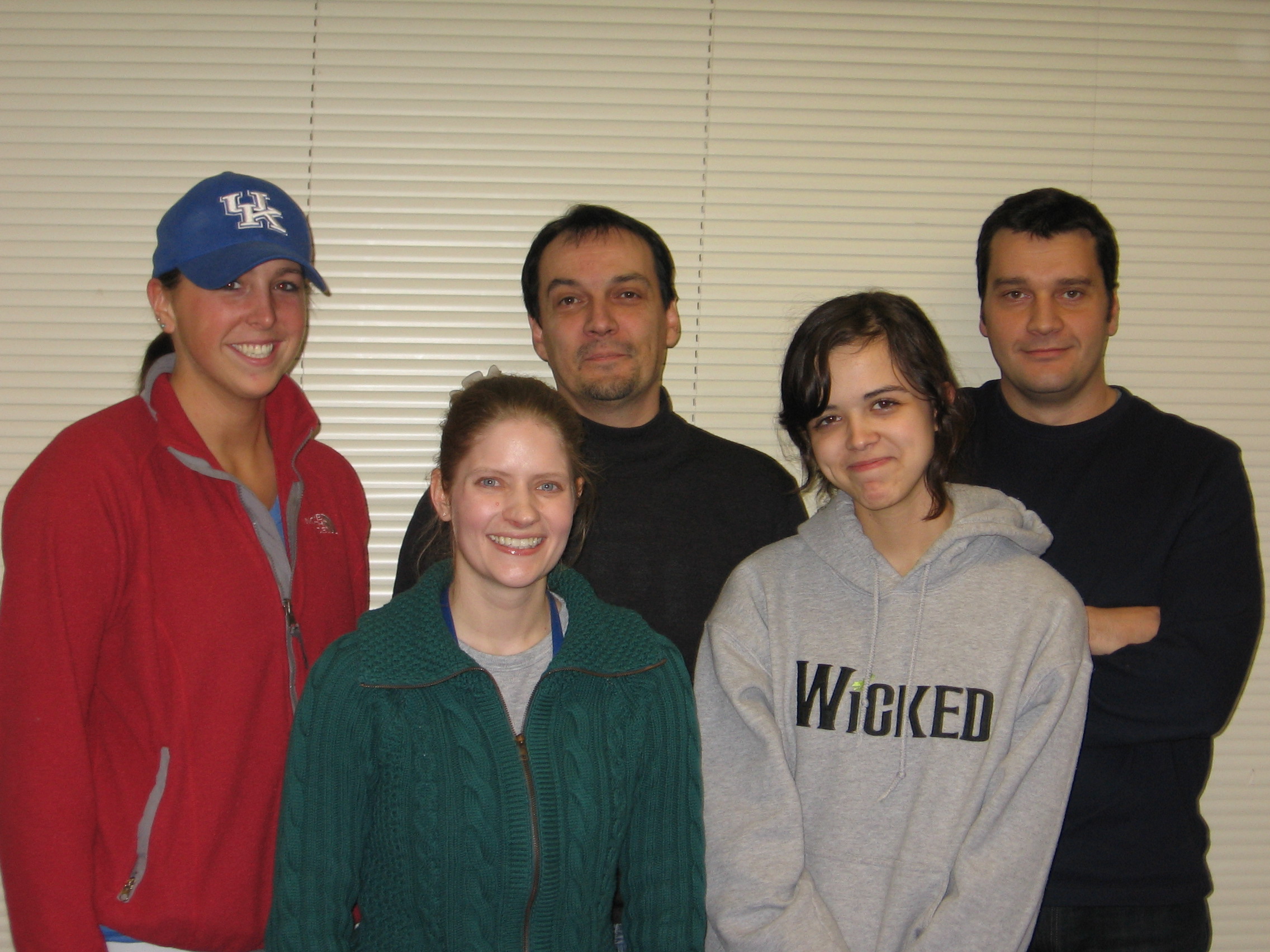
Meet the Lab:


.jpg)
.jpg)

.jpg)
Contact Us:
Our lab is located at the University of Kentucky, Department of Pharmacology and Nutritional Sciences, William R. Willard Medical Education Building, College of Medicine, UKMC MS-310.Dr. Olivier Thibault (Professor): othibau@uky.edu
Ruei-Lung Lin (Scientist): rueilung.lin@uky.edu
Sophiya Sims (Graduate Student): sophiyasims@uky.edu
Ting-Hsuan Lu (Postdoc Scholar): tinghsuan.lu@uky.edu
Leopoldine Galopin (Lab Tech): lbgalopin@uky.edu
Nicholas Wright (Lab Tech): nawrig2@uky.edu
Links:
Welcome to the Thibault Lab at the University of Kentucky!
Our lab primarily studies the effect of aging on astrocytic calcium dysregulation in a mouse model of amyloidosis (5xFAD). Previous work from our group focused on neuronal calcium dysregulation and associations with metabolic diseases (i.e., diabetes and hyperlipidemia) and the role of insulin in ameliorating cognitive decline and gait deficits. Our lab uses behavioral, electrophysiological, molecular, and fluorescent and 2P imaging techniques to interrogate neuronal and astrocytic function in health and disease. New projects currently under development include investigating the impact of overexpressing and knocking out insulin receptors in astrocytes and using metabolic nanosensors to assess astro- and neurovascular coupling in young and aged mice.
Students interested in research in the Department of Pharmacology and Nutritional Sciences at the University of Kentucky can contact the IBS Program.

Layers 2/3 somatosensory neurons expressing GCaMP8f flickering in response to animal ambulation (left). Astrocytic (GCaMP8f) and vascular (rhodamine dextran) responses to animal movement (right).
Techniques
In vivo 2-photon imaging of neurons and astrocytes in ambulating animals:
Ongoing work in the lab has been using state-of-the-art imaging techniques to assess neuronal and astrocytic
activity in young and aged mice. Currently, our work is focuses on using GCaMP to monitor alterations in
astrocytic calcium flux with movement. We also have been utiliting 2P imaging to evaluate metabolic health and
bioenergetic processess of live animals.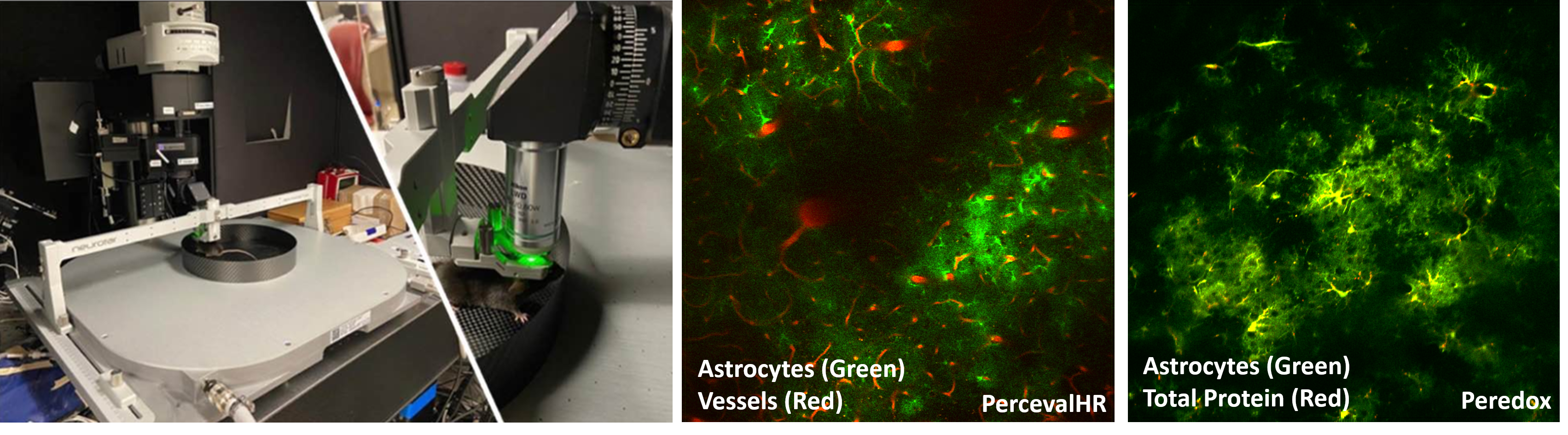
2P imaging of PercevalHR (ATP:ADP) and Peredox (NAD+:NADH) metabolic nanosensors in the awake animal.
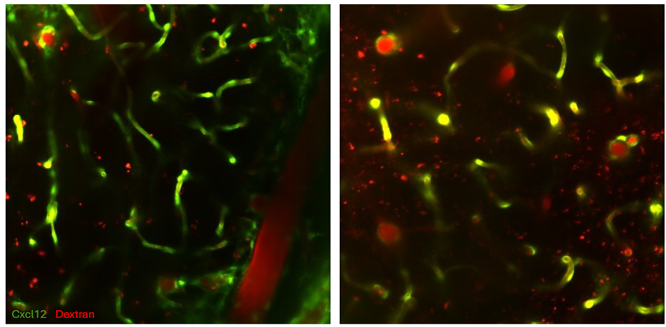
2P imaging of Cxcl12 (endothelial cell marker) and a fluorescent dextran (rhodamine) in the awake animal.
Fluorescent imaging:
We utilitze in vitro techniques (primary hippocampal neuronal and astrocytic culture) to image glucose
(2DG), ATP:ADP (PercevalHR), pH (pHrodo), and calcium (FURA) in live cells.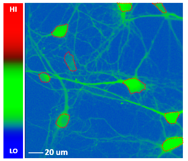
Fluorescent imaging of primary hippocampal cultures expressing PercevalHR.

A fluorescent glucose analog (2-NBDG) used to measure glucose utilization in neurons and astrocytes. Once inside the cell, 2-NBDG loses its fluorescence proportionally with the activation of glycolysis.
Immunohistochemistry and western blotting:
Using molecular approaches, we have characterized amyloid-beta deposition in our mouse model and evaluated astrocyte insulin receptor status on our in-house IR-KO and IR-OE mice using magnetic cell-separated western blotting techniques.
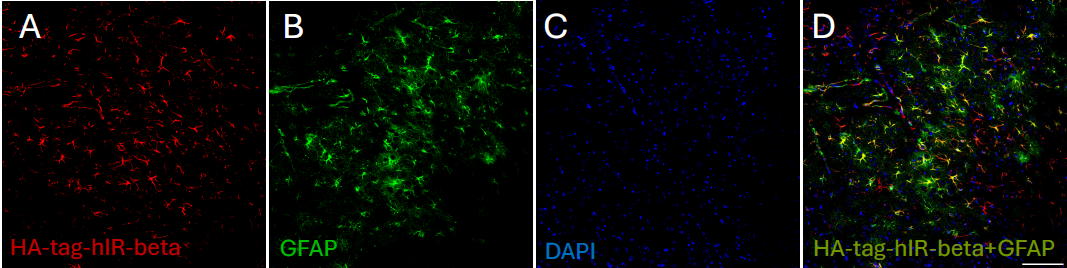
Double-label immunofluorescence of S1 in wildtype mice, showing HA-tagged-hIRbeta (A) and GFAP (B) immunoreactivity, with DAPI nuclear counterstain in blue fluorescence (C), as well as overlapping signals (D). Scale bar = 100 μm.
Vascular casting:
We use vascular casting to assess vessel density in our mouse model. Current investigations are evaluating the
effect of IRB-OE and IRB-KO on the angioarchitecture of the brain.
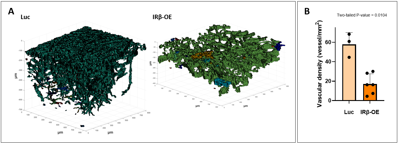
Representative images comparing the vascular density of control (Luciferase; [Luc]) and IR-B-overexpressed (OE) animals. A retroorbital injection of a fluorescein dextran was used to visualize local cerebrovasculature of layers 2/3 of S1 in control and IRβ-OE mice by 2P imaging. Scans were acquired at 5 µm slices to generate a 3D representation of the angioarchitecture. Quantification of vascular density reveals significant reductions in IRβ-OE mice.
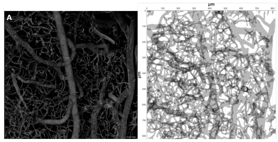
Example of vascular casting dervied from a mouse brain and quantification of vascular density using MATLAB.
Gait assessment:
We use a made-in-house ambulatory corridor to record and measure various gait parameters. Mice are permitted to
walk from one side of the corridor to the end and are assessed for variables such as stride length, stride time,
stride time deviance, steps per cm, and others.

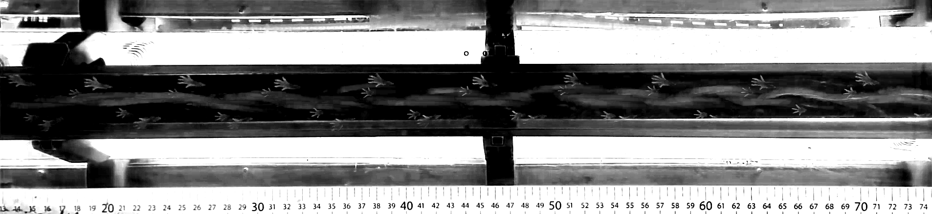
Mouse ambulation patterns from control (bottom) and 5xFAD (top) animals. Diseased animals display clear alterations in gait including shortened stride length and increased double support time. These measures reflect clinical manifestations of Alzheimer's Disease, where individuals may experience dysregulated gait, leading to falls.
Lab Research Focuses:
- Astro- and neurovascular coupling
- Brain aging and ADRDs
- Central impact of diabetes and metabolic disease
- Calcium signaling in neurons and astrocytes
- Neuronal and astrocytic network analyses
- Neurodegenerative diseases and amyloidosis
Recent Papers:
Lin RL, Sims SL, Wright NA, Galopin LB, Weiss BE, Kraner SD, Rhinehart JE, Lu TH, Sompol P, Norris CM, and Thibault O. (2025). Astrovascular decoupling in awake 5XFAD mice is associated with reduced astrocytic calcium. Alzheimer's & Dementia, Aug;21(8):e70564. DOI: 10.1002/alz.70564
Weiss BE, Gant JC, Lin RL, Gollihue JL, Kraner SD, Rucker EB, Katsumata Y, Jiang Y, Nelson PT, Wilcock DM, Sompol P, Thibault O, and Norris CM. (2025). Disrupted calcium dynamics in reactive astrocytes occur with endfeet-arteriole decoupling in an amyloid mouse model of Alzheimer's disease. Journal of Neuroscience, 2025 Oct 1;45(40):e0349252025 DOI: 10.1523/JNEUROSCI.0349-25.202
Parent R., Lin R.-L., Ouillette L., Glass E., Burns H., Smarsh A.T., Uhler M.D., Case S.L., Thibault O., and Murphy G (2025). The impact of overexpression of the mouse ortholog of CACNA1C on behavior and cortical dynamics. Biological Psychiatry: Global Open Science. DOI: 22;5(5):100537
Sims SL, Frazier HN, Case SL, Lin RL, Trosper JN, Vekaria HJ, Sullivan PG, Thibault O. (2024). Variable bioenergetic sensitivity of neurons and astrocytes to insulin and extracellular glucose. NPJ Metab Health Dis. 2(1):33. DOI: 10.1038/s44324-024-00037-y
Sami L. Case, Ruei-Lung Lin and Olivier Thibault (2023). Age- and sex-dependent alterations in primary somatosensory cortex neuronal calcium network dynamics during locomation. Aging Cell, DOI: 10.1111/acel.13898
Sami L. Case, Hilaree N, Frazier, Katie L. Anderson, Ruei-Lung Lin, and Olivier Thibault (2022). Falling short: The contribution of central insulin receptors to gair dysregulation in brain aging. Biomedicines. DOI: 10.3390/biomedicines10081923.