(16 of 16)
Junctional Epithelium
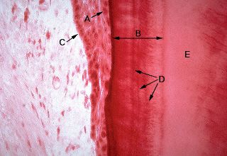
This image shows junctional epithelium near the cervix of the tooth. Electron microscopy would reveal hemidesmosomes between the epithelial cells (A) and the cementum (B), functioning to provide a form of attachment for the epithelium. Identify C, D and E.
Legend
|
A - epithelial cells |
D - resting lines |
Lecture Notes