TOPICS
- Home
- Challenges
- EIA Virus
- Transmission NEW!!
- Persistent Infection
- Pathogenesis
- Immune Response
- Diagnosis & Control
- Field Studies
- Publications NEW!!
- Links
- Contact and Credits
HOT TOPICS IN EIA RESEARCH
EIA Virus
General comments
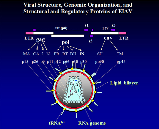
The virus that causes EIA is a relatively simple lentivirus and a close relative to the human immunodeficiency virus (HIV). It is simple in that its genomic organization includes the gag, gag-pol and env genes (that code for proteins in the virus particle) and only two additional short open reading frames or orfs (that code for regulatory functions). Please refer to the cartoon of the virus particle and the Figure (below) for more detailed information.
Although EIA was proven to be caused by a filterable virus in 1904, it was not until 1970 that an accurate diagnostic test was described. Once a diagnostic test was available, control of the disease was possible; testing became widely accepted by 1975. By the late 1970's much of the research for EIA became a low priority and few investigators remained active in the field. In Louisiana, our research expanded in two major areas -- applied research to define dynamics of transmission of the virus by insect vectors and basic research to define at the molecular level the "antigenic drift" of the EIA virus (EIAV) in horses with chronic disease. This latter phenomenon, first described by Japanese scientists, had been seen in other acute virus infections, e.g., influenza in humans, but had not been associated with persistent/chronic infections with viruses.
Infection-outcomes
In the United States today, EIA is most frequently diagnosed in apparently healthy equids because of required surveillance testing-moving interstate, going to congregation points and at transfer of ownership. This is quite different from the 1970's when most cases were found because of outbreaks of acute disease. Once an accurate laboratory diagnostic test was available we realized that the clinical cases represented only the tip of the iceberg.
The outcomes of infection with EIAV are determined by a number of host, virus and environmental factors. A virus strain that causes no disease in adult horses can kill early fetuses and foals who do not have the ability to mount immune responses against the virus. Likewise, a high dose of a virus strain that can kill an adult horse within 10-20 days of infection produces infection but no clinical disease in donkeys. The infected horse without clinical signs of EIA can develop signs of disease and shed dramatically higher amounts of virus after severe environmental stress or artificial doses of immunosuppressive drugs. See the figure below for graphic depiction of the clinical phases of EIA.
Body temperature (pink line) rises with exceptional virus replication and during the clinical bouts platelet counts often decrease to critical levels (blue line).  |
The only thing that we can say with certainty about infections with EIAV is that infection results in continuous virus presence in the equid host and that the clinical responses of the host through time are not predictable. Because all immune competent infected equids produce antibody against EIAV, usually within 60 days of infection, surveillance and control of the infection can be accomplished by testing horses for antibody.
Updated on: February 17, 2010
