
Basic and Translational Research Labs
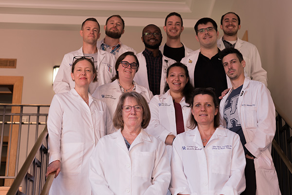 The basic science research labs occupy approximately 5,000 square feet on the 4th floor of the C.T. Wethington building, are overseen by seven College of Health Sciences’ faculty members, and are an integral part of the Center for Muscle Biology (CMB), directed by Dr. Charlotte Peterson. The CMB is home to 35 investigators, representing five colleges and 10 departments from across the UK campus with approximately $14 million in extramural research funding. The mission of the CMB is to support and integrate basic, clinical and translational research on muscle throughout the University of Kentucky. The goal is to understand mechanisms regulating muscle structure and function that impact overall health, to develop new strategies to improve physical performance and prevent frailty and the loss of functional independence following injury and in the face of chronic disease and aging.
 The laboratories of Drs. Esther Dupont-Versteegden, Christopher Fry and Charlotte Peterson are fully equipped with state-of-the-art equipment for cellular and molecular biologic analyses of rodent and human muscle tissues. The labs contain several tissue culture facilities for studying isolated primary muscle cells. Human tissue and primary muscle cell lines are stored in the CMB Human Muscle Bank, directed by Dr. Grace Walton. Shared equipment includes QuantStudio3 real time PCR detection system, BioRad ChemiDoc MP imaging system, and ZetaView (R) Nanoparticle Tracking Analysis system.
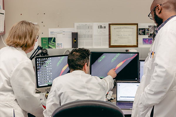
The wet labs are supported by a shared imaging facility that serves as the CMB Muscle Immunohistochemistry and Molecular Imaging Core (MIMIC), directed by Dr. Kate Kosmac. The facility includes three Micron cryostats, Zeiss AxioImager MI upright and AxioObserver D1 inverted fluorescent microscopes, each with fully automated stages for whole slide scanning and image stitching capabilities. The inverted scope is also equipped with an incubator system for live cell time lapse imaging. A third Olympus BX61VS upright fluorescent microscope has a fully automated stage which can hold up to 5 slides at once for whole cross-section imaging in batches. The MIMIC also provides access to MyoVision, an automated image analysis software program for quantifying muscle properties. 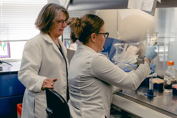
Dr. Tim Butterfield’s muscle mechanics lab contains animal treadmills, with integrated electromyography and high-speed camera systems to measure in-vivo kinematics and kinetics, and custom fabricated tissue loading devices and instrumentation for the measurement of passive and active mechanical properties of rodent muscle, in-vivo. The lab also contains custom fabricated mechanotherapy equipment (instrumented human and animal massage devices) for the application and quantification of external mechanical loads to human and animal skeletal muscle. A complete Aurora muscle physiology system overseen by Drs. Butterfield and Fry allows measurement of mechanical properties in isolated, whole muscle, including force-frequency, force-velocity, and force length, and permits additional measurements of fatigue, calcium sensitivity, rate of tension rise and recovery, passive muscle stiffness, and break length.
